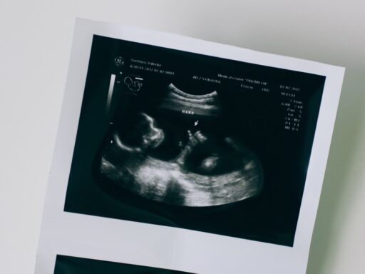
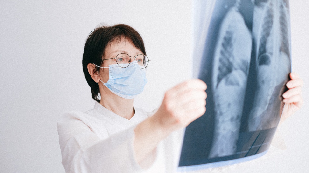

For children with chronic lung disease, doctors often face a difficult trade-off.
They need detailed images to understand whether the lungs are getting better, getting worse or responding to treatment. But some of the best-established imaging tools, particularly CT scans and X-rays, use ionizing radiation. One scan may involve a relatively small exposure, but children with lifelong conditions can require repeated imaging over many years.
Researchers at the University of Sheffield have developed a different approach.
Their technique combines magnetic resonance imaging with specially prepared xenon gas, allowing doctors to see not only the structure of a child’s lungs but also where air is actually moving through them. Because MRI uses magnetic fields and radio waves rather than ionizing radiation, the scans can potentially be repeated without adding radiation exposure.
The technology is already being used with children at Sheffield Children’s NHS Foundation Trust, where researchers say it can reveal signs of disease that conventional tests may miss.
That could make it particularly valuable for conditions such as cystic fibrosis, asthma and bronchiectasis.
Why Children’s Lungs Are Surprisingly Difficult to Scan
MRI is already one of medicine’s most important imaging technologies.
It works particularly well for organs and tissues containing lots of water. The brain, joints, muscles and many internal organs produce strong signals that MRI scanners can turn into detailed pictures.
The lungs present a very different challenge.
They are mostly filled with air.
Air produces relatively little of the conventional signal used by MRI, while constant breathing creates motion that can make imaging even more difficult. That is one reason chest X-rays and CT have traditionally remained so important in respiratory medicine.
The Sheffield researchers found a way to make the invisible air itself visible.
A child inhales a specially prepared form of xenon-129 gas before the scan.
The gas has been hyperpolarized, meaning its magnetic properties are temporarily enhanced enough to generate a strong signal inside the MRI scanner. The child holds his or her breath briefly while images are collected and then simply breathes the xenon back out.
Instead of seeing only what the lung looks like, doctors can see where the inhaled gas actually travels.
That changes what the scan can reveal.
The Scan Can Show Where Air Isn’t Reaching
Imagine looking at two lungs that appear structurally similar on an ordinary image.
One may function normally.
The other may contain small regions where narrowed or mucus-filled airways prevent air from reaching certain areas.
A conventional structural image may not always reveal those functional differences early.
Xenon MRI can.
The inhaled gas effectively creates a map of ventilation. Areas receiving air appear differently from regions where ventilation is impaired. The University of Sheffield says the technique can identify early functional abnormalities and monitor how those abnormalities change over time.
That may be particularly important for young children.
Children under five are often unable to reliably perform the standard breathing tests adults use to measure lung function. UK Research and Innovation notes that this makes early detection and monitoring especially challenging in pediatric respiratory medicine.
A scan requiring only a short breath hold could provide another source of information when conventional lung-function testing is difficult.
Cystic Fibrosis Could Be One of the Biggest Beneficiaries
Cystic fibrosis is a genetic condition that causes unusually thick mucus to accumulate in organs including the lungs.
Over time, that mucus can obstruct airways, encourage repeated infections and produce progressive lung damage.
Modern treatments have transformed outcomes for many people with cystic fibrosis, but monitoring remains essential because physicians need to know whether disease is progressing and whether particular therapies are working.
The Sheffield team says its lung MRI has identified ventilation abnormalities in children with cystic fibrosis even when conventional tests did not show obvious clinical lung disease.
That creates a potentially important advantage.
Earlier detection can give doctors more information before serious structural damage has developed.
Readers can explore the University of Sheffield’s work on pediatric lung MRI, where the researchers explain how the technology is already being integrated into clinical care.
The goal is not simply to produce prettier images.
It is to make treatment decisions earlier and with better evidence.
One Child’s Scan Changed Her Treatment Plan
UKRI describes the experience of a young patient named Zoe, who developed bronchiectasis after serious respiratory illness.
She had repeatedly needed intravenous antibiotics, and doctors were considering inserting a port into her chest to make future treatment easier.
Before that procedure went ahead, Zoe underwent two of the new MRI scans about two weeks apart.
The scans showed that the antibiotics were not producing a meaningful improvement in her lungs. Doctors therefore decided the port was unnecessary and changed the treatment approach toward daily physiotherapy to help clear mucus.
That example demonstrates another potential advantage of radiation-free imaging.
Doctors do not necessarily need to wait months before checking whether treatment is working.
They can potentially repeat imaging when clinically useful without accumulating the ionizing-radiation exposure associated with repeated CT examinations.
For chronic disease, that ability to look again can be almost as valuable as the first scan.
Why Avoiding Radiation Matters More in Children
CT remains an enormously valuable medical tool.
Its speed and spatial resolution make it indispensable in many situations, including emergencies and detailed evaluation of lung structure.
The new MRI technique does not mean CT suddenly becomes unsafe or unnecessary.
The issue is repeated exposure.
Children have longer expected lifespans ahead of them and developing tissues that can be more sensitive to ionizing radiation. Doctors therefore try to keep medical radiation exposure as low as reasonably achievable while still obtaining the information needed for care.
The UKRI overview of the Sheffield project emphasizes that repeated X-ray and CT imaging has to be balanced against those low-level radiation risks, especially when a child requires long-term monitoring.
MRI changes that equation because it does not use ionizing radiation at all.
That could allow clinicians to monitor some patients more frequently without facing the same cumulative exposure concern.
Xenon Can Reveal More Than Ventilation
The technique becomes even more interesting when xenon moves beyond the airways.
Some inhaled xenon dissolves through lung tissue and enters the bloodstream.
Researchers can detect those signals too.
That means advanced xenon MRI can potentially provide information about one of the lungs’ most fundamental jobs: transferring gases from inhaled air into the blood.
Combined with other MRI techniques, physicians may be able to evaluate ventilation, blood flow, tissue structure and gas exchange during the same broader examination.
That gives the technology a fundamentally different character from a simple anatomical scan.
It begins to resemble a functional map of the respiratory system.
Doctors can potentially ask not only, “What does this lung look like?”
They can ask, “Which parts are actually working properly?”
The Technology Took More Than 25 Years to Develop
Although the latest headlines make xenon MRI sound like a sudden breakthrough, Sheffield’s work has been developing for decades.
The university’s POLARIS MRI group and Insigneo Institute have spent more than 25 years developing pulmonary imaging methods, while collaborating closely with NHS hospitals and imaging manufacturers.
The technology also required more than inventing a new scan sequence.
Researchers needed equipment capable of hyperpolarizing xenon gas.
MRI hardware and software had to be adapted to detect it.
Clinical protocols had to be developed.
Regulatory approval had to be obtained.
The University of Sheffield says it led the technology through UK Medicines and Healthcare products Regulatory Agency approval and became the first institution to apply it clinically.
That long development process is why the current milestone matters.
A technology once confined largely to specialist research laboratories is beginning to move into actual patient care.
Existing MRI Scanners Could Potentially Be Adapted
Building an entirely new class of scanner for every hospital would make widespread adoption difficult.
The Sheffield team is taking a more practical approach.
Researchers have worked with GE HealthCare to develop hardware and software enabling compatible MRI systems to detect hyperpolarized gases. Sheffield says some of its lung-imaging protocols have already been incorporated into GE HealthCare systems, potentially making it easier for other hospitals to adopt the technology.
The research team is also investigating smaller, less expensive MRI systems that could eventually be deployed in more local clinical environments.
That could be important because a breakthrough available at only one specialist hospital has limited impact.
The long-term goal is making the technique practical enough to become part of ordinary respiratory care.
It Won’t Replace Every Chest X-Ray or CT Scan
This is where the excitement needs some perspective.
Pulmonary MRI is advancing quickly, but different imaging tools answer different questions.
CT remains exceptionally strong at resolving fine lung structures.
X-rays remain fast, inexpensive and widely available.
MRI is generally more expensive and technologically complicated, while hyperpolarized xenon requires specialized preparation and equipment.
The Sheffield researchers are therefore not suggesting that every child with a cough should immediately receive xenon MRI.
The strongest use cases are likely to involve children with chronic or complicated lung disease who need detailed functional information and repeated follow-up.
That is where eliminating radiation while obtaining additional physiological information becomes especially valuable.
The Biggest Breakthrough May Be Seeing Disease Before a Child Feels Worse
For families managing chronic respiratory disease, medicine often becomes reactive.
A child develops worsening symptoms.
Doctors perform tests.
Treatment changes.
Everyone waits to see what happens.
Radiation-free functional lung imaging offers the possibility of moving some of that process earlier.
Professor Jim Wild’s team says the technique can detect signs of disease before conventional lung-function tests and, in some situations, before abnormalities become apparent through conventional imaging.
If larger clinical studies continue supporting those findings, physicians may be able to identify deteriorating lung function before a child experiences a major decline.
That does not simply make the scan safer.
It could make respiratory care more proactive.
And for children who may need their lungs monitored for decades, that combination is particularly powerful.
The future of pediatric lung imaging may therefore involve something doctors have wanted for a long time: a way to look inside the lungs repeatedly, understand how they are functioning and intervene earlier without making radiation exposure part of the price of finding out.