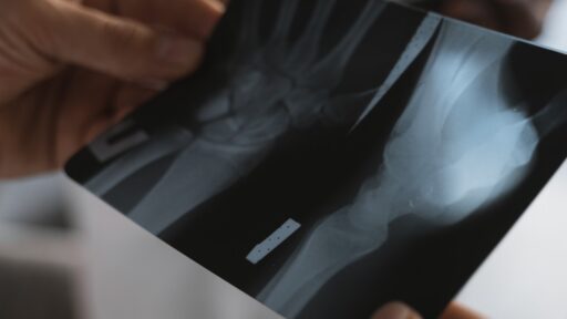
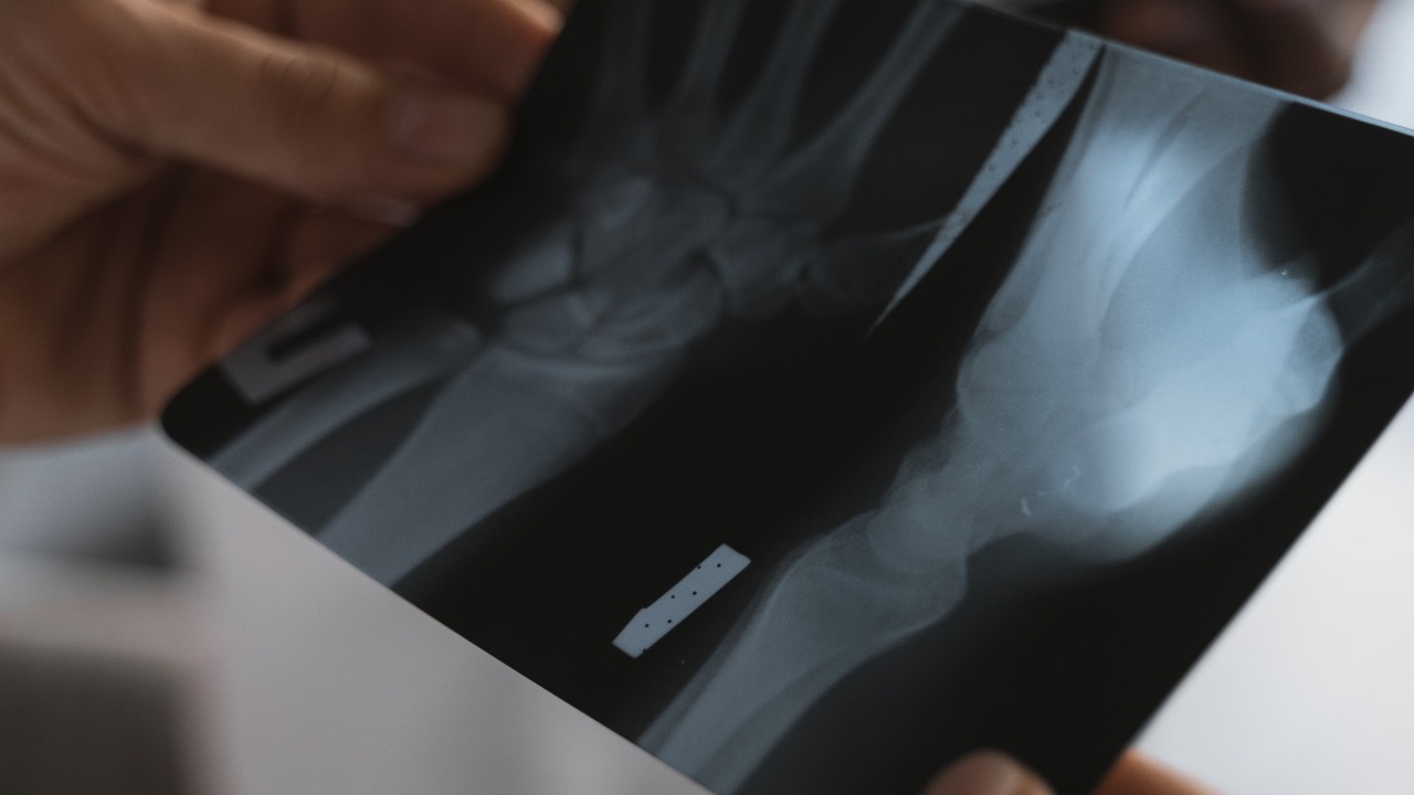

Astronauts have successfully captured the first diagnostic X-rays of human bodies during an orbital spaceflight, proving that compact radiography equipment can work in microgravity without direct support from medical specialists on Earth.
The images were taken during SpaceX’s private Fram2 mission using an ultraportable wireless X-ray system. Crew members with no professional radiology experience received only four hours of training before producing images of a hand, forearm, chest, abdomen and pelvis while orbiting Earth.
Three independent radiologists later examined the results and found that every in-flight image reached a diagnostically useful standard. The complete findings were published in the journal Radiology and summarized by the Radiological Society of North America.
The achievement could transform medical care during future missions to the Moon and Mars, where astronauts may be days or months away from a hospital and unable to return quickly after a serious injury.
The Historic Images Were Captured During Fram2
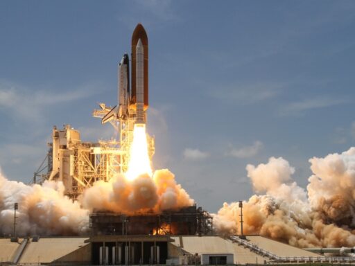
The breakthrough took place aboard Fram2, a privately funded SpaceX mission that launched on March 31, 2025.
The four-person crew travelled aboard the Crew Dragon spacecraft Resilience on the first crewed mission to enter a polar orbit around Earth. Fram2 flew at an altitude of approximately 425 to 450 kilometres and remained in space for three days and 14 hours before returning on April 4.
The mission’s unusual orbit allowed the crew to pass over Earth’s polar regions, but it also served as a platform for multiple scientific and medical experiments. SpaceX provides an overview of the flight through its official Fram2 mission page.
Three crew members were trained to operate the X-ray system before launch. None were professional radiographers, and their training lasted just four hours.
They first captured comparison images on Earth, then repeated the procedures during the flight. After returning, additional images were collected using the same protocols so researchers could compare results from before, during and after the mission.
Astronauts X-Rayed Several Parts of the Body
The crew produced in-orbit X-rays of a hand, forearm, abdomen, pelvis and chest.
They also imaged a calibration object and a smartwatch to test whether the device could examine equipment as well as human anatomy.
The images were recorded digitally and transmitted immediately to an onboard computer. This meant the crew could review them without developing film or sending physical material back to Earth.
The RSNA’s detailed report states that the portable system operated without real-time ground assistance. That level of independence is essential for future deep-space crews, who may experience long communication delays or temporary loss of contact with Earth.
The ability to capture images of central body regions was particularly important. A hand or arm can be positioned relatively easily, but chest, abdominal and pelvic X-rays require more precise alignment between the patient, detector and radiation source.
The Images Were Good Enough for Medical Diagnosis
Three independent radiologists evaluated all the X-rays.
They compared overall image quality, spatial resolution, contrast resolution and patient positioning. The researchers found no meaningful difference in overall quality, contrast or spatial resolution between the images captured before launch and those obtained in orbit.
Positioning scores were lower for some chest, abdominal and pelvic images, which was expected because both the equipment and the subject were floating in microgravity. Even so, every in-flight image reached a diagnostic standard.
That means the images contained enough useful information for doctors to identify conditions such as fractures and assess some internal medical problems.
Coverage from Space.com noted that the Earth-based images were generally easier to position, but the orbital results remained sufficiently clear for practical diagnosis.
The result directly challenges the long-standing assumption that useful radiographs would be too difficult to produce inside a constantly moving spacecraft.
Why X-Rays Had Never Been Used This Way in Space
Astronauts have lived and worked in space for decades, but ultrasound has remained the main medical imaging tool available during missions.
Ultrasound is compact and does not expose the patient to ionizing radiation. It can examine internal organs, muscles, blood vessels and other soft tissues.
However, it requires substantial operator training. The person holding the probe must apply it at the correct angle and position while recognizing the anatomy displayed on the screen.
X-rays can be faster and more useful for diagnosing broken bones, certain chest problems and some types of internal injury. They are also easier for specialists on Earth to interpret once a clear image has been captured.
Traditional X-ray systems were unsuitable for spacecraft because they were large, power-hungry and sensitive to movement. Launch vibration and landing forces could damage their internal components, while floating patients and equipment increased the likelihood of blurred or badly aligned images.
The miniaturization of radiography equipment has changed that calculation. The Fram2 system used an ultraportable wireless generator and digital detector that could fit inside a spacecraft already carrying a crew, supplies and scientific equipment.
Microgravity Created a Major Positioning Challenge
On Earth, gravity helps keep the patient, X-ray source and detector in a stable position.
Inside a spacecraft, every object floats unless it is secured. Even a small movement while the image is being captured can reduce sharpness or cause the wrong part of the body to appear on the detector.
The Fram2 crew used available supports and positioning techniques, but later recommended adding stronger clamps and mounting systems to future versions of the equipment.
Those additions could help secure the detector, generator and astronaut in a repeatable arrangement.
This lesson may become especially important on lunar or Martian missions. A spacecraft in transit would operate in microgravity, while the Moon and Mars would provide partial gravity that is weaker than Earth’s.
Medical devices may therefore need flexible mounting systems capable of operating under several different gravitational conditions.
Radiation Exposure Remained Within Clinical Levels
Sending an additional source of radiation into space creates an obvious concern.
Astronauts are already exposed to more cosmic radiation than people on Earth because they have less protection from the atmosphere and magnetic field. Unnecessary medical radiation should therefore be minimized.
Researchers estimated that exposure from the Fram2 X-rays was no greater than that associated with standard clinical radiography on Earth.
That does not mean repeated imaging would be risk-free. Every examination would still need a medical justification, particularly during a long mission when cumulative radiation exposure is already elevated.
Future procedures will need clear guidelines explaining when an X-ray is necessary, which body parts should be examined and how to use the lowest radiation dose capable of producing a useful image.
The study’s authors said additional research is required to establish examination standards, reference images and interpretation protocols for people living in space.
X-Rays Could Diagnose More Than Broken Bones
Fractures are the most obvious use, particularly after a fall, equipment accident or hard landing.
However, radiography could assist with several other medical emergencies. Chest X-rays may help doctors investigate lung problems, fluid buildup or complications after trauma. Abdominal imaging could reveal some foreign objects, bowel problems or equipment-related injuries.
A portable system could also help determine whether an injured astronaut can continue working or requires evacuation.
That decision becomes increasingly important as missions move farther from Earth. Returning from the International Space Station can take hours or days depending on spacecraft readiness. A lunar crew may be several days away, while a Mars mission could be months from emergency care.
The American College of Radiology’s overview explains that diagnostic radiology uses X-rays and other imaging methods to identify injuries and diseases without exploratory surgery. That same benefit becomes even more valuable when surgery, specialists and full hospital facilities are unavailable.
The Equipment Could Inspect Spacesuits and Electronics
The experiment also demonstrated that portable radiography could examine nonmedical objects.
The crew successfully X-rayed a smartwatch and a calibration object. Researchers believe similar systems could look inside spacesuits, electronics and mechanical components without taking them apart.
That could help astronauts locate damaged wiring, cracked connectors or hidden structural failures during a mission.
A spacecraft carries limited replacement parts, and disassembling essential equipment can create additional risks. X-ray inspection could reveal where a failure has occurred before the crew begins repairs.
The technology may also assist with malfunctioning satellites or be mounted on lunar rovers to study rocks and soil. The RSNA research summary identifies hardware inspection and lunar exploration as possible future applications.
The Machine Survived Launch and Re-entry
The portable system had to endure vibration during launch, orbital operation and the forces of atmospheric re-entry and ocean recovery.
After the mission, researchers found superficial damage to the machine’s exterior. Its internal hardware and X-ray output were unaffected.
That survival is important because medical equipment intended for deep-space travel must remain reliable after months of vibration, temperature changes and radiation exposure.
A device that works perfectly in a hospital may fail when subjected to rocket launch forces. Fram2 therefore tested not only the quality of the images but also the physical durability of commercially available equipment.
Future models will likely need stronger housings, better attachment points and simplified controls designed specifically for gloved or fatigued crew members.
The Technology Could Also Help Remote Communities on Earth
Portable X-rays developed for space may provide significant benefits far from orbit.
Compact systems can be powered in areas without full hospital infrastructure and may transmit digital images to specialists through a laptop, tablet or smartphone.
They could support rural clinics, disaster-response teams, ambulances and temporary medical facilities. Patients in isolated communities might receive an initial diagnosis without travelling several hours to a large hospital.
The devices could also assist after earthquakes, floods or conflicts where conventional radiology departments are damaged or inaccessible.
Researchers emphasized that portable systems are already used at sporting events and in low-resource settings. Spaceflight testing may now encourage the development of smaller, more durable and easier-to-operate versions.
A Major Step Toward Independent Space Healthcare
The Fram2 experiment does not mean every spacecraft will immediately carry an X-ray machine.
Medical planners must still consider equipment mass, power use, radiation exposure, training requirements and the limited space available inside crew vehicles.
Image interpretation is another challenge. Astronauts may capture the X-ray themselves, but a qualified radiologist would normally assess it from Earth. Future Mars missions could face communication delays of several minutes in each direction, making real-time consultation impossible.
Artificial intelligence may eventually assist with preliminary interpretation, although automated systems would require extensive testing before they could guide treatment in a high-risk environment.
Even with those limitations, the first successful orbital radiographs represent a major change in space medicine.
For more than 40 years, ultrasound was the only dependable medical imaging method used during human spaceflight. Fram2 has now shown that minimally trained crew members can operate portable X-ray equipment, produce diagnostically useful images and inspect both people and hardware while travelling around Earth.
As missions move toward permanent lunar bases and eventual journeys to Mars, astronauts will need greater medical independence. Carrying a compact X-ray system could help crews understand injuries quickly, make better treatment decisions and avoid unnecessary emergency returns.
The milestone is therefore about more than a set of historic images. It is an early demonstration of the medical tools humans will need to live and work far beyond Earth.