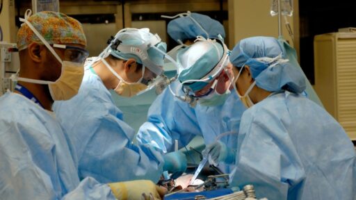
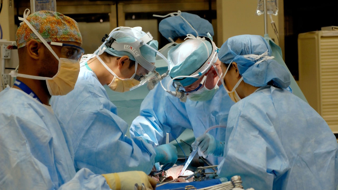

Surgeons preparing for a complex brain operation can now step inside a three-dimensional reconstruction of the patient’s anatomy before entering the operating theatre.
Using virtual reality, augmented reality and mixed-reality headsets, medical teams can examine tumours, aneurysms, blood vessels and delicate brain structures from different angles. They can enlarge the anatomy, remove virtual layers, mark a possible surgical route and rehearse difficult stages of an operation without placing the patient at risk.
The technology turns conventional MRI and CT scans into interactive, patient-specific models. Instead of planning entirely from a sequence of flat images, surgeons can explore a digital version of the individual they are about to treat.
A recent BBC Tech Now feature followed medical teams using these immersive environments to prepare for high-risk procedures. The technology was also used to show a patient exactly where an abnormality was located and how the surgical team intended to reach it, making the consultation more detailed and potentially less intimidating.
From Flat Medical Scans to a Virtual Patient
CT and MRI scans already provide surgeons with detailed information, but they are normally reviewed as two-dimensional slices on a computer monitor. The surgeon must mentally combine those slices to understand the three-dimensional relationship between a tumour, nearby blood vessels, nerves and healthy tissue.
Immersive software performs part of that reconstruction digitally.
Platforms developed by companies such as XRlabs can convert standard patient scans into an interactive anatomical model, sometimes described as a digital twin. Wearing a mixed-reality headset, the surgeon can view the model as a hologram positioned within the room, walk around it and inspect structures from perspectives that would be difficult to reproduce on a conventional screen.
The original scan data remains essential. The headset does not discover anatomy that was invisible to the scanner. Its purpose is to present the information spatially, making complicated relationships easier to understand.
This can be especially valuable in neurosurgery, where a difference of only a few millimetres may affect whether a surgical route approaches a critical artery, an optic pathway or a region controlling speech and movement.
Surgeons Can Rehearse the Actual Case
Traditional surgical simulation usually teaches a standard procedure on a generic virtual patient. Patient-specific immersive planning goes further by allowing the team to practise using the anatomy of the person scheduled for surgery.
A surgeon can assess several routes toward a tumour, estimate the position and size of the required opening, inspect where major vessels pass and anticipate which structures may restrict access. The rehearsal can also expose uncertainties that require further imaging or discussion before the operation begins.
Imperial College Healthcare NHS Trust has introduced mixed-reality technology into neurosurgical planning and patient consultations. Its teams use the system to transform conventional scans into high-resolution, three-dimensional images that clinicians can examine collaboratively.
In February 2025, neurosurgeons in London and Washington also used the XRlabs platform to review the same patient-specific cases together in a shared mixed-reality environment. The collaboration allowed specialists separated by thousands of miles to enter the same virtual workspace, point to anatomical structures and discuss surgical strategies as though they were standing beside the same model.
That possibility is important for rare or highly complex conditions. A hospital may not have a local surgeon with extensive experience in one unusual procedure, but immersive collaboration could allow a distant specialist to contribute to the planning.
The Technology Can Make Patient Consent More Meaningful
Immersive surgical planning is not intended only for doctors.
Patients are commonly shown MRI or CT images during consultations, but interpreting those images can be difficult without medical training. A collection of grey anatomical slices may not clearly communicate where a tumour lies, why surgery is dangerous or what the surgeon plans to do.
A patient-specific three-dimensional model can make the explanation more intuitive. The clinician can identify the abnormality, show nearby structures and demonstrate the proposed route through the skull or surrounding tissue.
The patient featured by BBC said seeing her own brain in mixed reality was reassuring rather than frightening. The experience helped her understand the operation and participate more fully in the consent process.
A 2025 feasibility study involving patient-specific brain imaging also found that mixed-reality consultation was practical and could improve understanding of neurological anatomy and proposed treatment. However, the researchers stressed that larger studies are needed before firm conclusions can be made about clinical benefits.
Informed consent involves more than obtaining a signature. A patient needs to understand the purpose of the procedure, its main risks, possible alternatives and what may happen without treatment. Immersive visualisation could strengthen that conversation when it is used carefully and accompanied by a clear explanation.
Medical Trainees Can Practise Without Risking a Patient
Surgical education has traditionally relied on observation, supervised participation, cadavers, physical models and simulation devices. Each method remains valuable, but access can be limited.
A rare operation may not occur during a trainee’s rotation. Cadaver laboratories are expensive and cannot reproduce every living response. Opportunities inside an operating theatre must also balance education with patient safety and time pressure.
Virtual environments can be repeated as often as necessary. A trainee can practise the same step, review an error and attempt the procedure again without causing bleeding or permanent injury.
A 2026 meta-analysis found that virtual-reality training had a significant positive effect on surgical performance in operating-room and closely related tasks, although the included studies varied considerably in their methods and quality.
Another systematic review found that VR-based robotic-surgery training improved task performance compared with receiving no additional training. Performance was broadly comparable with conventional simulator training in the studies analysed, although more research was needed to establish how consistently the skills transferred to real operations.
The benefit is therefore not that a headset can instantly create an experienced surgeon. It provides another controlled environment in which technical decisions, hand movements and spatial understanding can be developed before those abilities are required in a real emergency.
AI Could Make Virtual Rehearsals More Personalised
Artificial intelligence is increasingly being integrated into immersive medical platforms.
AI-assisted software can help segment medical scans by separating bones, vessels, nerves, tumours and soft tissues. Automating this process can reduce the time required to create a usable three-dimensional model.
A 2026 study of patient-specific spinal-surgery simulation used computer-vision methods to combine CT and MRI scans and automatically generate anatomical models. The system produced models in approximately two and a half minutes per case and allowed users to rehearse decompression procedures inside a virtual operating room. Surgeons and trainees reported improved spatial understanding and confidence, although the research remained an early technical and educational evaluation rather than proof of better patient outcomes.
Future systems may compare several surgical approaches, identify structures at risk and provide performance feedback after a rehearsal. They could also analyse tool movement, decision timing and errors to create personalised training plans.
AI recommendations would still require clinical oversight. Anatomical segmentation can be inaccurate, and an error in a virtual model could misrepresent the relationship between important structures.
Mixed Reality May Eventually Enter the Operating Theatre
Preoperative planning is only one possible application. Some augmented-reality systems are being developed to display guidance during surgery.
A headset could theoretically place a patient’s scan over the real surgical field, giving the surgeon a view similar to a navigation map. Important vessels, tumour boundaries or planned entry points could appear in the surgeon’s line of sight without requiring constant attention to a separate monitor.
Commercial systems such as Brainlab’s Mixed Reality Viewer already allow clinicians to review vascular anatomy, estimate the location of an opening and assess possible clip positions before cranial procedures.
Intraoperative use is more difficult because the virtual image must remain precisely aligned with the patient. Tissue can move after an incision, the brain can shift during surgery, and even a small registration error could make an overlay misleading.
For that reason, immersive guidance cannot be treated as an independent replacement for established surgical navigation, imaging or direct anatomical judgment.
The Evidence Is Promising but Not Complete
Immersive technology frequently improves spatial understanding, learner confidence and planning experience. Evidence that it consistently improves major patient outcomes is less developed.
A systematic review of patient-specific immersive VR found that surgeons generally reported a better preoperative-planning experience, but evidence for improvements in complications, recovery or other short-term clinical outcomes remained limited.
A broader 2026 review found that virtual planning prompted surgeons to modify their intended approach in roughly one-third to one-half of cases across some of the included studies. That suggests the models can influence decisions, but a changed plan is not automatically a better plan. Controlled studies must still determine whether the change reduces risk or improves recovery.
Cost, staff training, software compatibility, headset discomfort and the time required to prepare models can also restrict adoption. Patient scan data must be protected, while software used for clinical decisions may require medical-device approval and formal quality controls.
Immersive Technology Will Support Surgeons, Not Replace Them
The strongest role for immersive technology is not autonomous surgery. It is giving human clinicians a clearer way to understand anatomy, communicate with colleagues and prepare for difficult decisions.
A headset cannot reproduce the full reality of living tissue. It cannot completely simulate bleeding, unexpected anatomical variation or the pressure of responding when a patient’s condition changes suddenly.
It can, however, make the first encounter with a complex case happen before the patient reaches the operating theatre.
For medical trainees, that means practising dangerous steps without real-world consequences. For experienced surgeons, it means exploring the individual anatomy behind the scans. For patients, it may provide a clearer explanation of what will happen and why.
The technology is still developing, and its impact on patient outcomes requires stronger evidence. Yet its direction is clear: lifesaving surgery is becoming something a medical team can explore, discuss and rehearse in three dimensions before making the first incision.